Cardiac Review 3D — 3D printing and surgical planning
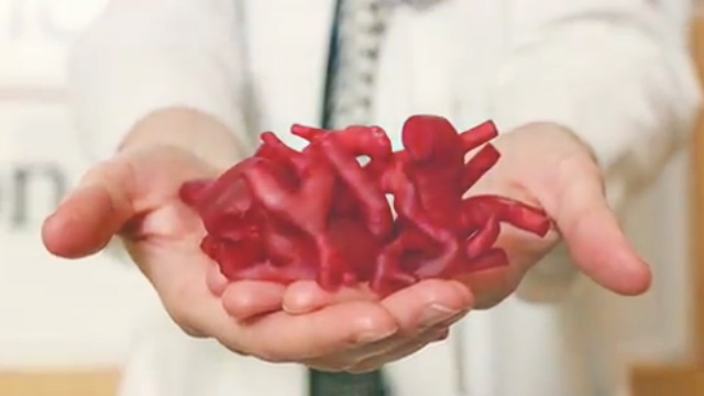
In fall 2014, we kicked off a collaboration with a group of scientists and physicians at the Sheikh Zayed Institute for Pediatric Surgical Innovation at Children’s National Hospital in Washington, DC. We have been developing software with Dr. Axel Krieger and Dr. Laura Olivieri in support of their research into using 3D surface models to help improve communication between cardiac imagers and cardiovascular surgeons. This group is already known for innovative work investigating the application of 3D printing to surgical planning for congenital heart defect repair. This video shows how they turn patient scans into handheld physical objects that accurately reproduce patient-specific cardiac anatomy.
Our application is called Cardiac Review 3D. It’s designed to facilitate the collaborative review and annotation of surface models generated from medical scans, such as computed tomography (CT), magnetic resonance imaging (MRI) and echocardiography (echo). It’s a natural extension of our partner’s 3D printing research, providing a digital platform to review and share the surface models that they are currently sending to be physically printed.
A number of applications already exist to load and visualize 3D surface models, but our software was designed from the ground-up for this specific research purpose, providing fluid and natural interactions, simple labeling/annotation tools, and PDF report generation. In addition to mouse-and-keyboard inputs, the application was also designed for a portable touch-based interface (check out “The Platform” below) so that it can be easily carried throughout the hospital.
This post overviews our design process from initial specifications to the final software.
The Specs
In discussions with our research partners, we settled on a set of specifications:
- Windows software to load STL (Stereolithography) files
- Portable platform that can be easily carried around a hospital
- Touch-driven and mouse/keyboard interface
- Interactive labeling and measurements
- Screenshot feature to capture images from the interface
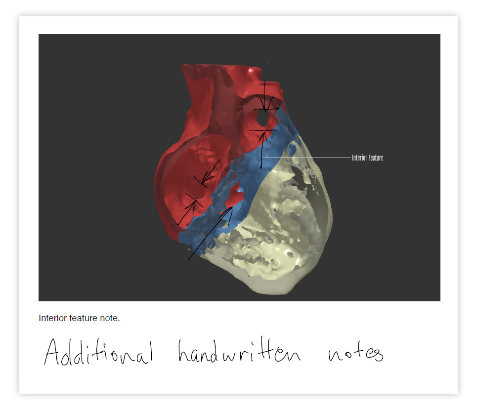
- Report export feature to combine screenshots and notes into a single PDF document
Our goal: Make loading, reviewing, and annotating surface models as simple as possible.
The Mockup
We use Balsamiq to produce mockups for all of our software projects. It allows us quickly iterate the user interface and program flow together with our clients. Changing the mockup is quick and simple, whereas changing finished software is difficult and costly.
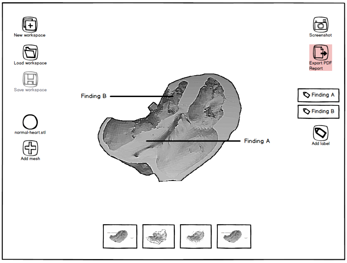
The hand-drawn style prevents the design process from getting bogged down with low-level details like button color or font choices. After a few iterations, the result is an agreed-upon mockup that our team codes against, exported as a hyperlinked PDF document that feels very much like using the final software. Click here to download our actual PDF mockup for this project.
The Platform
We selected the Microsoft Surface Pro 3 because it hit the right balance of portability and performance. The detachable keyboard means that users can annotate the surface model, then later share and review their work with the computer detached in tablet mode. We decided to develop for Windows in order to simplify file management and streamline the connection with existing servers and IT infrastructure.
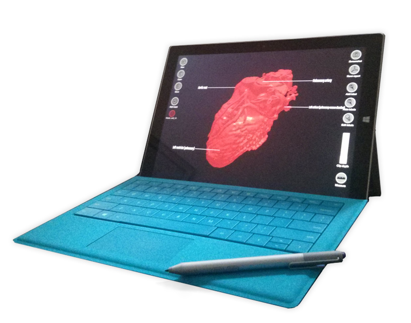
The Surface has enough graphics horsepower to run the application smoothly with the typical ~5-30MB models (although she does get warm!). The pen input is a nice bonus as it can be used to draw additional markup or handwritten text directly onto the exported PDF using the native Windows 8 document viewer. The model in the image below was downloaded from the excellent NIH 3D Print Exchange website.
The Result
We delivered the first version of the application to our scientific partners at Children’s National, who will incorporate it in a larger surgical planning study. We also announced and showcased the software at the recent NIH Science in 3D conference on January 20th, 2015. Video clips from this event can be found below (including words from Dr. Krieger from Children’s National and Matt Irwin, who is a principal here at Indicated). Stay tuned for updates about the next steps for this application.
The cardiac case study featured in the Surface 3 image and in Dr. Krieger’s presentation was published here: Olivieri L, Krieger A, Chen MY, Kim P, Kanter JP. 3D Heart Model Guides Complex Stent Angioplasty of Pulmonary Venous Baffle Obstruction in a Mustard Repair of D-TGA. Int J Cardiol. 2014 Jan 8. We would like to thank Dr. Dilip Nath, Dr. Lillian Su, and the Children’s National Board of Visitors (BOV) for funding part of this work.